3D Imaging in Zygomatic Bone Fracture Treatment
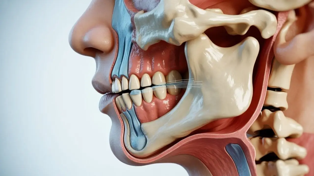
3D imaging has become an important tool in the assessment of zygomatic bone fracture cases. This technology provides detailed views of facial bone structures that traditional two-dimensional radiographs may not fully capture.
Patients who experience facial trauma often require precise evaluation to understand the extent of injury. 3D imaging helps visualize fracture lines, bone displacement, and surrounding soft tissue relationships in a single scan.
The following sections explain how 3D imaging supports diagnosis, planning, and follow-up in zygomatic bone fracture management while emphasizing that every case needs individual professional assessment.
What Is a Zygomatic Bone Fracture?
A zygomatic bone fracture involves the cheekbone area of the facial skeleton. This type of injury commonly results from direct impact during accidents, falls, or sports activities.
Symptoms may include swelling, bruising, changes in facial contour, and difficulty with jaw movement. Accurate identification of fracture patterns is essential before any treatment approach is considered.
How 3D Imaging Supports Diagnosis
3D imaging, typically obtained through cone-beam computed tomography or medical CT, creates multiplanar and volumetric reconstructions of the zygomatic bone fracture. These images allow clearer visualization of fracture segments and their spatial relationships.
- Fracture displacement can be measured in three dimensions.
- Orbital floor involvement and maxillary sinus extension become easier to evaluate.
- Associated injuries to the temporomandibular joint or alveolar bone can be identified simultaneously.
Treatment Planning Using 3D Data
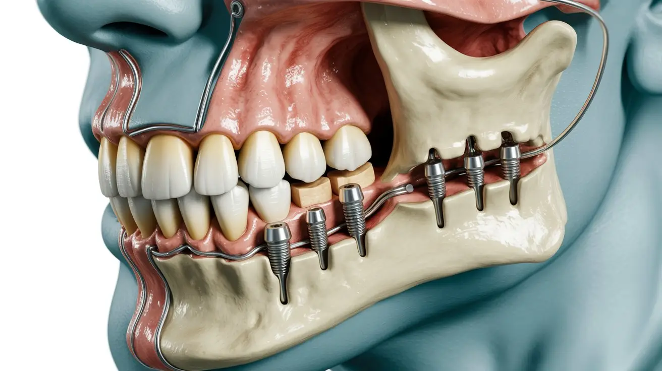
Once a zygomatic bone fracture is confirmed, 3D imaging assists surgeons in deciding between conservative observation and surgical intervention. Virtual models help simulate reduction and fixation options.
| Aspect | 2D Radiographs | 3D Imaging |
|---|---|---|
| Fracture detail | Limited overlap views | Clear spatial orientation |
| Planning accuracy | Basic estimation | Precise segment positioning |
| Soft tissue relation | Poorly visible | Better assessment possible |
| Follow-up comparison | Difficult alignment | Standardized 3D registration |
Post-Treatment Evaluation and Follow-Up
After initial management of a zygomatic bone fracture, repeat 3D scans may be used to verify bone alignment and healing progress. This helps detect complications such as malunion or persistent displacement at an early stage.
Regular monitoring remains important because healing responses vary between individuals. Personal clinical examination combined with imaging findings guides further decisions.
Sık Sorulan Sorular
Is 3D imaging required for every zygomatic bone fracture?
Not every zygomatic bone fracture needs 3D imaging. The decision depends on clinical findings, suspected complexity, and the need for surgical planning. A qualified professional determines the most appropriate imaging method after examination.
How does 3D imaging differ from standard X-rays in facial trauma?
Standard X-rays provide flat images, while 3D imaging offers multi-angle and volumetric data. This additional detail can improve understanding of fracture displacement and adjacent structure involvement in zygomatic bone fracture cases.
Can 3D imaging show soft tissue damage around the fracture?
Yes, 3D scans can reveal relationships between bone fragments and surrounding soft tissues. However, additional imaging modalities may still be required for detailed evaluation of muscles, nerves, or blood vessels.
Is radiation exposure higher with 3D imaging?
3D imaging generally involves more radiation than plain radiographs. Dose levels vary by device and protocol, so the benefit-risk balance is assessed individually for each patient.
Does 3D imaging guarantee better treatment outcomes?
3D imaging supports more informed planning but does not guarantee outcomes. Final results depend on fracture type, patient health, surgical technique, and healing factors that require professional follow-up.
When is the best time to perform 3D imaging after facial injury?
Timing depends on swelling, patient stability, and clinical priorities. In many zygomatic bone fracture situations, imaging is performed once acute swelling subsides to allow clearer interpretation.
Sonuç
3D imaging offers valuable information for evaluating and planning the management of zygomatic bone fracture injuries. Its use should always be guided by individual clinical findings and professional judgment rather than routine application.
Expert Information
This content is prepared for general informational purposes in oral, dental and maxillofacial surgery. Prof. Dr. Celal Çandırlı’s over 20 years of professional experience, with training at England NHS Poole Hospital and Newcastle Freeman Hospital, contributes to addressing the topic within a scientific and clinical framework.
This content is for general informational purposes only and does not replace professional medical advice. Individuals with facial trauma or zygomatic bone fracture concerns should consult a qualified healthcare professional for personalized evaluation.