The bone zygomatic—widely known as the cheekbone—is one of the most essential facial bones that shapes midface aesthetics, forms the lateral orbital wall, supports mastication, and protects the eye. Understanding the detailed anatomy, function, symptoms of fractures, and clinical significance of the bone zygomatic is crucial in maxillofacial surgery and facial trauma management.
Bone Zygomatic Anatomyfacial
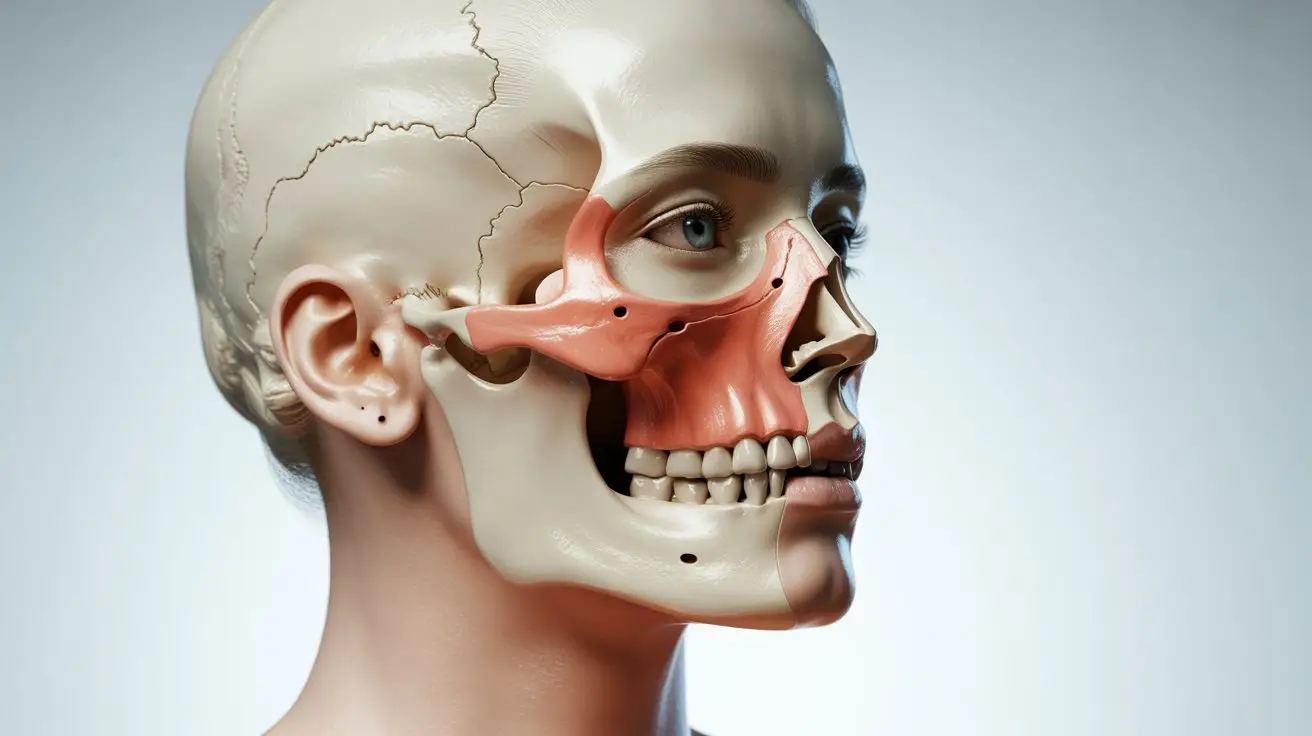
The bone zygomatic forms the prominence of the cheek and contributes to facial contour and skeletal strength. It articulates with multiple craniofacial structures, making it a pivotal element of facial harmony. Location and Structural Composition The zygomatic bone is located in the lateral midface region. It is composed of cortical and cancellous bone, providing a balance of rigidity and lightweight structural support.
Articulations of the Zygomatic Bone
The bone zygomatic connects with:
Frontal bone
Maxilla
Temporal bone
Sphenoid bone
Key Anatomical Landmarks
Zygomaticofacial foramen
Zygomaticotemporal foramen
Lateral orbital rim
Muscle attachment surfaces
Zygomatic Bone Frontal Process
Zygomatic Bone Frontal Process
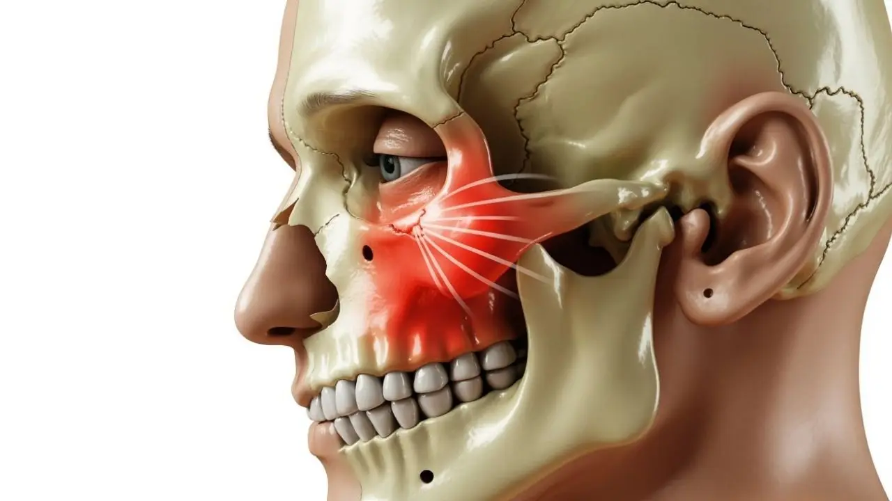
The frontal process of the bone zygomatic extends upward and forms the lateral orbital margin. This region plays a vital protective role for the eye. Trauma to this process may affect orbital stability, eyelid function, or infraorbital nerve sensation—common concerns in facial fractures.
Zygomatic Bone Muscle Attachments
Several facial expression and mastication muscles attach directly to the bone zygomatic, making it a key structural point in both movement and facial aesthetics. The most important of these are the zygomaticus major, which elevates the corners of the mouth and creates the smiling expression, and the zygomaticus minor, responsible for lifting the upper lip. Additionally, the lateral fibers of the orbicularis oculi muscle attach to this region and support eyelid movements, while the masseter muscle connects through the zygomatic arch to generate significant chewing force. Together, these muscles highlight the functional and expressive importance of the bone zygomatic in daily facial activity.
Functional Implications of the Bone Zygomatic
Injuries involving the zygomatic bone may lead to facial asymmetry, weakened bite force, or loss of expression control—particularly affecting the smile.
Zygomatic Bone Pain
Zygomatic Bone Pain
Pain in the bone zygomatic region may result from fractures, nerve compression, sinus disorders, dental issues, or post-surgical healing.
Common causes of pain in the bone zygomatic region include a variety of structural and neurological factors. A zygomatic fracture is one of the most frequent sources, often accompanied by swelling, bruising, and facial asymmetry. Infraorbital nerve irritation may also lead to sharp or radiating pain beneath the eye, while temporomandibular joint disorders can create referred discomfort in the cheek area. Infections such as sinusitis may produce pressure-like pain around the zygomatic bone, and trauma or post-operative swelling can further contribute to localized tenderness and sensitivity. Understanding these potential causes is essential for accurate diagnosis and effective treatment.
Zygomatic Bone CT Anatomy
CT scans are the most accurate method to evaluate the bone zygomatic for fractures, displacement, and orbital floor involvement.
At Celal Candırlı Clinic, advanced maxillofacial diagnostics begin with Dr. Celal Candırlı’s innovative 3D Imagination Technique, a modern approach that allows highly detailed visualization of the bone zygomatic and surrounding facial structures. This technique enhances diagnostic accuracy by combining three-dimensional imaging principles with clinical expertise, enabling precise assessment of fractures, orbital involvement, and midface symmetry. As a result, patients benefit from more accurate treatment planning, improved aesthetic outcomes, and a deeper understanding of their facial anatomy before any surgical intervention.
CT Imaging Shows
Zygomatic arch displacement
Orbital wall fractures
Infraorbital foramen damage
Zygomaticomaxillary junction integrity
CT Indications
Trauma with facial deformity
Unilateral cheek flattening
Double vision
Pre-surgical planning
Bone Zygomatic And Its Function
The bone zygomatic maintains facial contour, protects the eye, supports chewing, and plays a critical role in facial appearance and biomechanics. The primary functions of the bone zygomatic play a crucial role in both facial structure and biomechanics. It is responsible for creating cheek prominence and midface projection, which significantly influence overall facial aesthetics. Additionally, the zygomatic bone provides essential orbital support, helping protect the eye from lateral trauma. Another key function is the distribution of masticatory forces through its connection to the masseter muscle, allowing efficient chewing mechanics. Beyond these structural roles, the bone zygomatic also contributes to facial expression control through its interaction with muscles involved in smiling and upper lip movement, making it an integral element of both function and appearance.
ZMC fractures involve the bone zygomatic, orbital floor, and maxilla. Early diagnosis improves long-term outcomes.
Diagnosis & Evaluation
Diagnosis and evaluation of conditions involving the bone zygomatic require a combination of clinical assessment and advanced imaging methods. The process begins with a detailed clinical symmetry assessment, allowing the surgeon to identify flattening, contour loss, or deviations caused by trauma. A CT scan of the facial bones is then performed to visualize fractures, displacements, and orbital wall involvement with high precision.
Sensory testing of the infraorbital nerve is also essential, as nerve irritation or damage can cause numbness beneath the eye. Additionally, evaluation of eye movement helps determine whether the zygomatic injury has affected the orbital structures, which may lead to double vision or restricted ocular mobility. Together, these steps ensure a comprehensive and accurate assessment of the bone zygomatic before planning treatment.
Treatment Options
Treatment options for injuries involving the bone zygomatic depend on the severity of the fracture, degree of displacement, and functional impact on the orbital or masticatory structures. Closed reduction may be performed in cases where the bone is only mildly displaced, allowing the surgeon to reposition the zygomatic bone without making large incisions.
More complex injuries often require open reduction and internal fixation (ORIF),a method in which small titanium plates and screws are used to stabilize the bone zygomatic and restore facial symmetry. In situations where the fracture affects the orbital floor or wall, orbital reconstruction may be necessary to protect the eye, prevent double vision, and maintain proper orbital support. Each technique aims to restore both the functional and aesthetic
integrity of the zygomatic region.
Zygomatic Bone Surgical Approaches
Surgical correction restores the bone zygomatic structure, symmetry, and orbital protection—often using minimal-incision techniques.
Common Surgical Methods
Transconjunctival incision
Lateral eyebrow incision
Intraoral (vestibular) access
Gillies temporal approach for arch reduction
Aesthetic Importance of the Bone Zygomatic
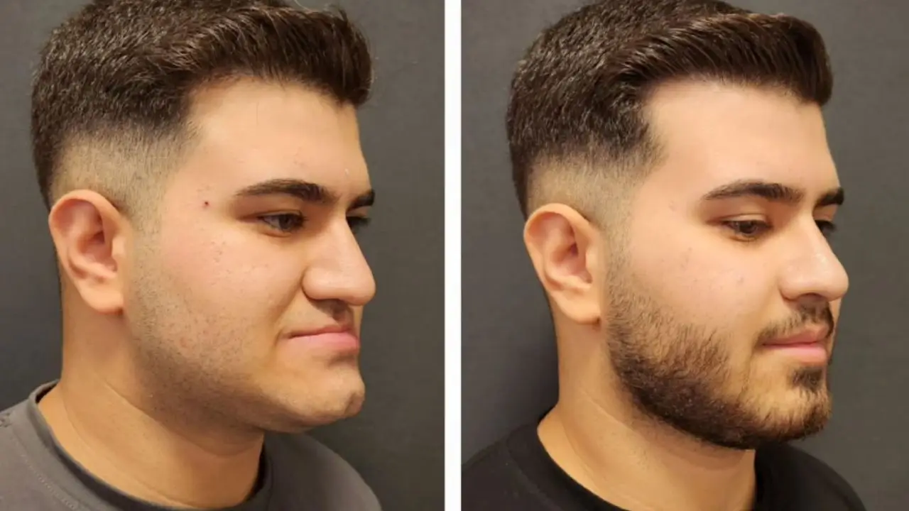
The bone zygomatic defines midface projection and youthful facial contour, making it a key focus in cosmetic facial surgery.
Related Cosmetic Procedures
Zygoma reduction surgery
Cheek augmentation
Facial contouring
Frequently Asked Questions About Bone Zygomatic
The bone zygomatic is crucial for facial aesthetics, protection, and function. Understanding its anatomy, fracture symptoms, and surgical approaches is essential for accurate diagnosis and treatment. Patients experiencing trauma, persistent pain, or facial asymmetry should seek evaluation by a facial plastic surgeon or maxillofacial expert.
What is a zygomatic bone?
The zygomatic bone, often referred to as the cheekbone, is a key structural component of the midface. It forms the lateral wall of the orbit, contributes to facial symmetry, and creates the prominence of the cheeks. The bone zygomatic plays a vital role in both aesthetics and function, supporting chewing muscles and protecting orbital structures.
Is zygomatic bone attractive?
Yes. Prominent and well-defined zygomatic bones are widely associated with facial attractiveness and youthfulness. High cheekbones create stronger facial definition, enhance midface projection, and contribute to a balanced facial profile. For this reason, the bone zygomatic is often a focus in cosmetic procedures such as cheek augmentation and facial contouring.
Why is my zygomatic bone hurting?
Pain in the bone zygomatic area can be caused by trauma, sinus inflammation, nerve irritation, dental issues, or temporomandibular joint disorders. In some cases, the discomfort may indicate a zygomatic fracture or post-operative swelling. Persistent cheekbone pain should be assessed by a specialist to rule out structural injuries or nerve involvement.
What is another name for a zygomatic bone?
Another name for the zygomatic bone is the malar bone. This term is commonly used in anatomy, maxillofacial surgery, and aesthetic medicine, especially when referring to cheek augmentation or midface reconstruction.
How do you know if you have a zygomatic bone injury?
A bone zygomatic injury often presents with symptoms such as cheek flattening, swelling, bruising around the eyes, numbness under the eye due to infraorbital nerve irritation, pain while chewing, restricted jaw movement, or double vision. CT imaging is the most accurate method to confirm a zygomatic fracture or displacement.
Is cheekbone pain serious?
Cheekbone pain can be serious if it is caused by a fracture, nerve compression, sinus infection, or orbital injury. If the pain is accompanied by swelling, asymmetry, vision changes, or numbness, evaluation of the bone zygomatic is essential to prevent long-term functional or aesthetic complications.
Can you feel your zygomatic bones?
Yes. The bone zygomatic is superficial and easily palpable beneath the skin. It forms the outer contour of the cheek, so any changes in its shape—such as flattening, protrusion, or asymmetry—can often be felt by touch.
What is the rarest face type?
The rarest face type is generally considered to be the diamond-shaped face, characterized by narrow forehead and jawline with pronounced zygomatic bones. This facial structure relies heavily on the natural prominence of the bone zygomatic.
What’s the strongest bone in your face?
The strongest bone in the face is the mandible (lower jaw), but the bone zygomatic is also highly durable and crucial for structural support. It withstands significant masticatory forces through its connection to the masseter muscle and helps distribute impact across the midface.